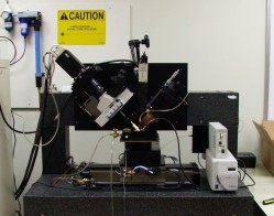
 | A full view of the Knife-Edge Scanning Microscope. Note the upgraded knife assembly on the right (also see the close-up shot right below).
[AVI Movie] (10.4MB) [AVI Movie: different encoding] (2.6MB)
NEW 7/18/2005
|
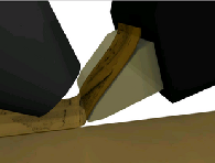
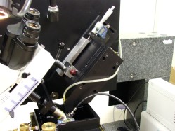 | Redesigned knife assembly and light source (right) for improved chatter resistence.
NEW 7/18/2005
|
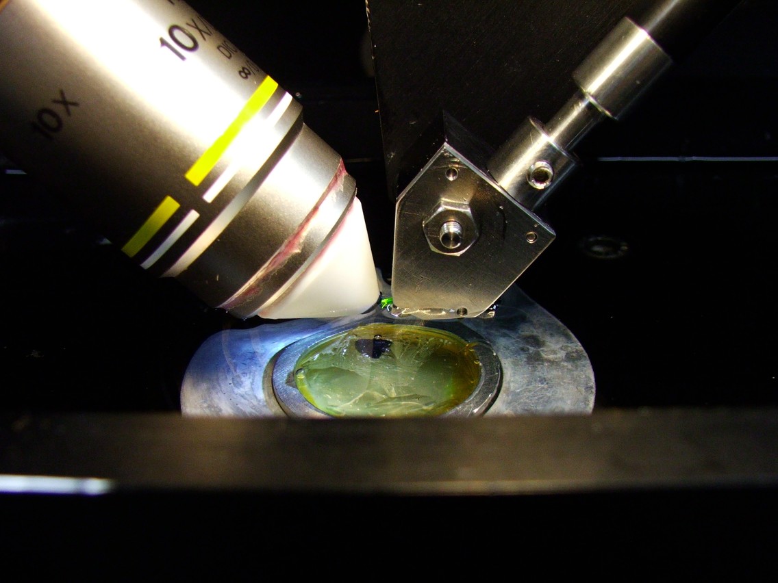
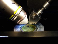 | Cutting session: Microscope objective (left) and the diamond knife (right) in place for cutting. The specimen (block in the center) and the knife are fully submerged.
[AVI Movie] (9.9MB) [AVI Movie: different encoding] (1.57MB)
High-res version:[AVI Movie] (30MB) [AVI Movie: different encoding] (5.71MB)
NEW 7/18/2005
|
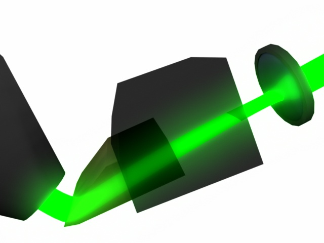
 | An illustration of how the diamond knife provides illumination. The objective is shown on the left and the white light illumination and the knife assembly are shown to the right.
|
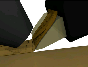
 | An illustration of how the diamond knife cuts the tissue. The objective is shown on the left and the light source and the knife assembly are shown to the right.
|
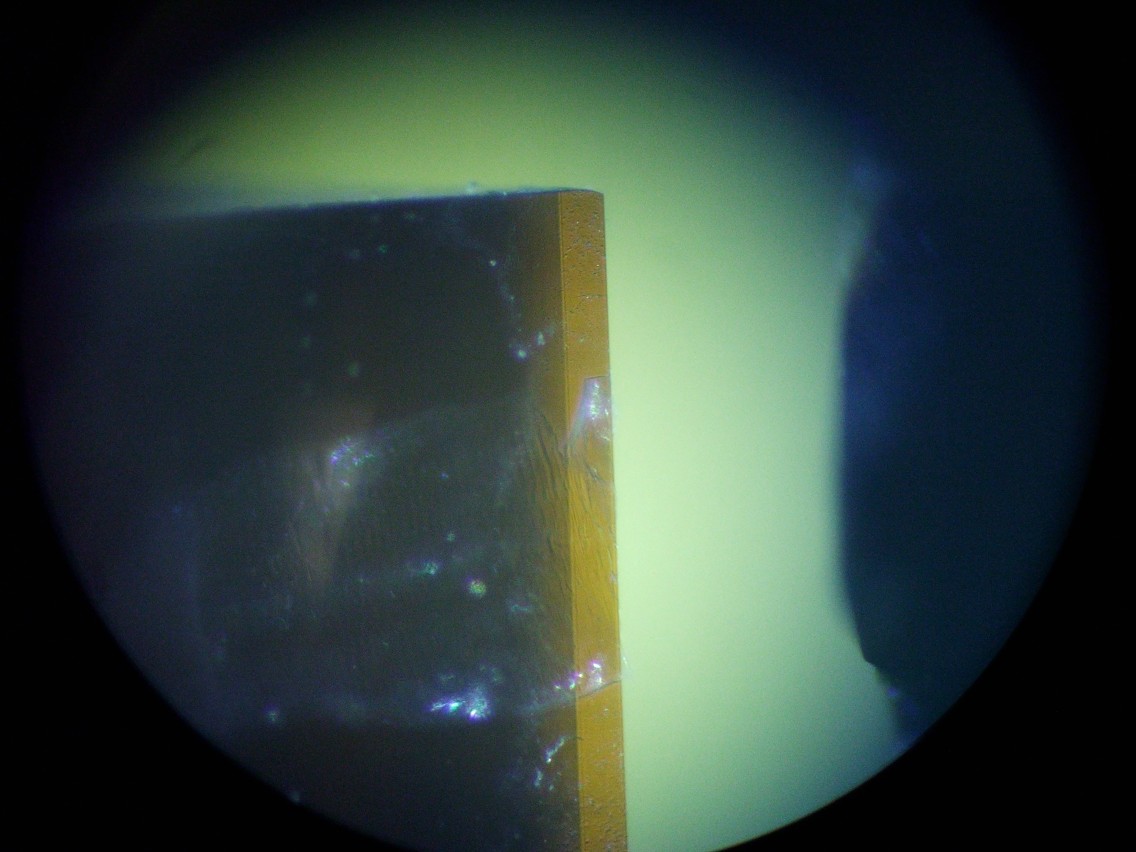
 | The tip of the diamond knife as seen through the observation eyepiece. The translucent tissue just cut off is shown on top of the diamond knife (left). The osmium-stained mouse brain can be seen below to the right. Note that this movie is shown in real time!
[Low-res AVI Movie] (17.9MB) [Low-res AVI Movie: different encoding] (4.26MB)
[High-res AVI Movie] (37MB) [High-res AVI Movie: different encoding] (6.5MB)
NEW 7/18/2005
|
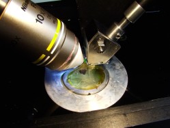 | Closeup of KESM optics and diamond knife assembly. The green glowing part is the diamond knife, and the black blob below is the osmium-stained mouse brain embedded in LR-White.
NEW 7/18/2005
|
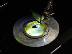 | Closeup view of the submerged mouse brain (osmium stained) with the light turned on on the diamond knife (the green glowing part).
NEW 7/18/2005
|