Click on the image to see a PDF version (for zooming in)
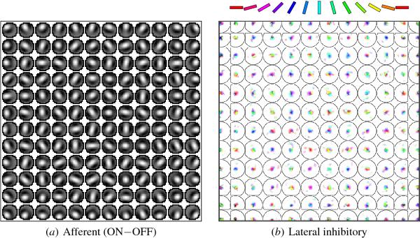
Fig. 5.8. Self-organized afferent and lateral weights across
V1. This plot shows the range of afferent and lateral weights
developed by the neurons in the orientation map, by plotting them for
every 12th neuron horizontally and vertically, using the conventions
introduced in Figure 5.7. (a) A number of two- and three-lobed
receptive fields exist with strong orientation preferences. Some
neurons, however, have ring-shaped RFs and respond to all directions
equally. (b) These neurons receive lateral inhibitory connections from
all nearby neurons, but from distant neurons only if they have similar
OR preferences and are located along the preferred orientation. The
lateral excitatory connections of each neuron (not shown) come from
all nearby neighbors, and thus are all nearly circular.
|

