


BNL News
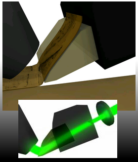
Diamond knife/illuminator: The diamond knife is used both as a cutting instrument and as an illuminating component of KESM. [More images] [More nuggets] |
- Portraits of the MIND: Book page on amazon
Our whole-brain vasculature data set has been featured in the recently published book "Portraits of the MIND" by Carl Schoonover!
- CNS*2010 Workshop on Highthroughput Microscopy: CNS*2010 Workshop on High-throughput 3D microscopy and high-performance computing for multi-scale modeling and simulation of large-scale neuronal circuits
An archive of presentation slides from the minisymposium is now available: Workshop Archive.
- Whole brain Nissl scanned!:
In January 2010, we have achieved another landmark event. We scanned an entire Nissl-stained mouse brain at 0.6 um x 0.7 um x 1.0 um resolution. The results are reported in Society for Neuroscience abstracts.
- NSF Grant Awarded:
Our KESM whole-brain Golgi and vasculature data set can now be made pubilc! We received an informatics grant from NSF to make our data sets available to the public through an intuitive web-based interface.
- Soc. for Neurosci Minisymposium: Society for Neuroscience Minisymposium on High-throughput Microscopy Microscopy and Computational/Theoretical Challenges in the Analysis of Neural Circuit Structure
An archive of presentation slides from the minisymposium is now available: Minisymposium archive.
- Gallery page updated: BNL gallery page (data fly-throughs and 3D reconstruction)
The gallery page has been updated, with new whole-brain data sets and visualization. See Data fly-throughs and 3D reconstruction.
- Publications page updated: BNL publications page
The publications page has been updated to include latest publications between 2006 and 2008.
- Whole mouse brain scanned (Golgi):
The KESM project is celebrating a landmark event: The first full scan of a Golgi-stained mouse brain at a sub-micrometer resolution (0.6 um x 0.7 um x 1.0 um voxel size). The Golgi stain labels full neuronal morphology for approximately 1% of the entire neuronal population. Sectioning/imaging started 7/7/08 and ended 8/5/08 (8 hours/day, 5 days/week, with intermittant down-time for maintenance)
- Whole mouse brain scanned (India ink):
The KESM project achieved a major breakthrough. The first full scan of a India-ink-stained mouse brain at a sub-micrometer resolution (0.6 um x 0.7 um x 1.0 um voxel size). India ink, perfused through the vascular network of the mouse brain, labels the full network of blood vessels. Sectioning/imaging started 4/11/08 and ended 4/23/08 (8 hours/day, 5 days/week, with intermittant down-time for maintenance)
- Former director Dr. Bruce H. McCormick passed away: Departmental Notice
In memory of Bruce H. McCormick (1928-2007): Dr. McCormick, founding director of our lab, passed away on 11/30. Dr. McCormick had a life-long dedication to interdisciplinary research, with a great vision and drive for brain networks research. His insights, enthusiasm, collegiality, and good humor will be missed greatly by everyone.
- New data flythroughs uploaded: Gallery
Flythrough movies of new Nissl data have been uploaded (rat cortex).
- New data flythroughs uploaded: Gallery
Flythrough movies of new vascular data have been uploaded (mouse spinal cord).
- New data flythroughs uploaded: Gallery
Flythrough movies of new Nissl+India ink data have been uploaded (mouse olfactory bulb).
- KESM 1.5 technical spec uploaded: Publications
A technical report detailing KESM 1.5 optics and cameras have been uploaded in the pulications page.
- Multiscale Modeling webpage: Multiscale Modeling webpage
The main MSM consortium web page, including project descriptions for all awarded projects.
- KESM automation software completed:
The fully automated sectioning and imaging software for KESM has been completed.
- Gallery movies: Codec update: Gallery
The AVI files in Gallery have been converted to play on most players without a specific codec.
- New KESM photos and movies: KESM Gallery
New photos and videos of KESM operation have been added. See the entries marked New 7/18/05
- New Nissl and Golgi data and reconstruction results: Data page (Gallery); Reconstruction page (Gallery)
New Golgi and Nissl dataset and reconstructions have been added. The reconstruction include microvascular data obtained from Nissl-stained tissues. See the entries marked New 7/7/05
- New reconstruction results: Gallery page
Two new animations of reconstruction results have been added. One for KESM Golgi and one for EM data. See the entries marked New 3/11/05
- Patent awarded to KESM: US patent was awarded to KESM.
See 2004 news below.
- Two New MS theses available: Download the PDF files in the publications page.
Two new MS theses on segmentation and construction of polymerized data sets are available. See the publications link above: by Prathyusha Aragonda and by Purna Doddapaneni.
- Patent awarded to KESM: US patent was awarded to KESM.
NEWS RELEASE: Patent issued for the Knife-Edge Scanning Microscope (KESM)
The United States Patent and Trademark Office (USPTO) issued a patent (US 6,744,572) on June 1, 2004, covering the principles underlying the knife-edge scanning microscope, an instrument for imaging a specimen in three dimensions at submicron resolution. The patent, "System and Method for Imaging an Object", is held by the Technology Licensing Office, Texas A&M University, with Bruce H. McCormick, inventor. In the instrument, the specimen is physically sectioned and imaged concurrently, unlike the age-old tradition in histology, for example, where these two processes are separately performed. This concurrency of serial sectioning and imaging allows the instrument to maintain three-dimensional registration of the stack of images so generated.
In one implementation of the Knife-Edge Scanning Microscope, the specimen (for example, a whole mouse brain) is embedded in a plastic block and mounted atop a three-axis precision positioning stage. A custom diamond knife, rigidly mounted to a massive granite bridge overhanging the stage, cuts consecutive serial thin sections from the block. A light source illuminates the tissue at the diamond knife tip with a strip of intense illumination reflected from the beveled knife edge. The diamond knife performs two distinct functions: as an optical prism in the collimation system of the microscope and as the tool for physical sectioning (a key patent claim). A microscope objective, aligned perpendicular to the top surface of the knife, images the illuminated narrow band of tissue in the newly cut section just beyond the knife edge, prior to subsequent deformation of the tissue ribbon. This image is digitally encoded by a high-sensitivity line-scan camera, and then stored in a cluster computer.
June 1, 2004 -- BNL - NSF REU students join the BNL: Research Experience for Undergraduates summer program begins.
Two undergraduate students joined the BNL this week to be part of the various projects in the lab. These students are supported by the NSF REU program, awarded to the computer science department at Texas A&M University.
Contact us | Home
© Copyright 2004 Brain Networks Laboratory | Department of Computer Science
Dwight Look College of Engineering | Texas A&M University | Texas Engineering Experiment Station

