Click on the image to see a PDF version (for zooming in)
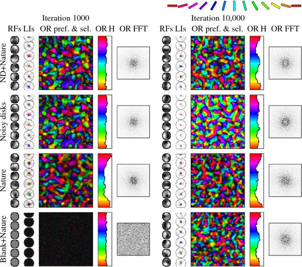
Fig. 9.6. Effect of prenatal and postnatal training on orientation
maps. The different rows illustrate how the prenatal training
phase affects the final selforganized maps. The state of each network
at iteration 1000 is shown on the left half, and the final state at
iteration 10,000 on the right half. In the "ND+Nature" simulation (the
same as in Figures 9.1 and 9.3), postnatal training makes more neurons
sensitive to horizontal and vertical contours and more selective in
general. However, the overall map shape remains similar, as found
experimentally in animals (Chapman et al. 1996; compare individual
orientation patches between pairs of maps on the top row). However,
even without any prenatal training (bottom row), or when the network
is trained with natural images also prenatally (third row), HLISSOM
develops a qualitatively similar final map. In these cases, its
organization depends only on the properties of the natural images, not
on the internally generated patterns under genetic
control. Conversely, even when natural images are replaced by
internally generated ones in postnatal training (second row),
orientation maps still develop. However, they are not a good match to
the visual environment: For example, the orientation histogram is
essentially flat. These results suggests that prenatal training is
useful mostly because it allows animals to have a functional visual
system already at birth, forming a robust starting point for further
development. Postnatal training, on the other hand, allows the animal
to adapt to the actual visual environment.
|

