Click on the image to see a PDF version (for zooming in)
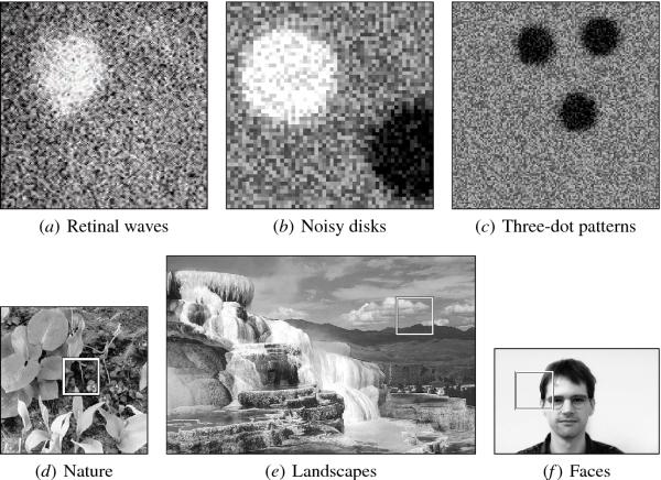
Fig. 8.4. Internally generated and environmental input
patterns. The three top images depict prenatal input patterns on
the retina and the PGO pathway in gray scale from black to white (low
to high). (a) A sample retinal wave pattern from the ferret (see also
Figure 1.2) is used to motivate the actual patterns in HLISSOM
experiments. (b) The "noisy disk" representation of retinal waves is
used to organize the orientation map prenatally. A light disk models
activity in the ON channel and a dark disk that in the OFF
channel. (c) A PGO activity configuration of three dark noisy disks,
corresponding to the two eyes and the nose/mouth area, is proposed to
underlie prenatal development of face preferences. The three bottom
images are samples of visual inputs, including those of (d) nature,
(e) landscapes, and (f) faces. Randomly located retina-size segments
(such as those shown by white squares) are used to train and test V1,
and full face images to train and test the FSA, measuring how the
variation in postnatal training affects the orientation map and how
the face preferences develop postnatally. Sources: (a) Feller et
al. (1996), (d) Shouval et al. (1996, 1997), (e) National Park Service
(1995), (f) Achermann (1995), copyright 1995 by University of Bern.
|

