Click on the image to see a PDF version (for zooming in)
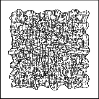
Fig. 5.11. Retinotopic organization of the orientation map. The
center of gravity of the afferent weights of every second neuron was
calculated and plotted in the retinal space, and those of neighboring
neurons connected with lines (as in Figure 4.8). The overall
organization of the map is an evenly spaced grid with local
distortions. These distortions result from mapping both orientation
and retinal position smoothly into the same two-dimensional surface;
such distortions have been found experimentally on animal maps as well
(Das and Gilbert 1997).
|

