Click on the image to see a PDF version (for zooming in)
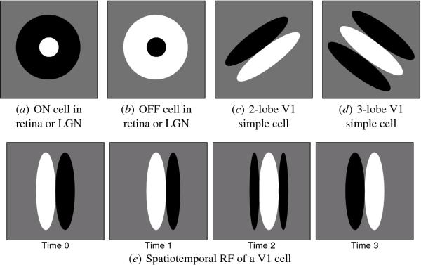
Fig. 2.2. Receptive field types in retina, LGN and V1. Each
diagram shows a receptive field on the retina for one neuron. Areas of
the retina where light spots excite this neuron are plotted in white
(ON areas), areas where dark spots excite it are plotted in black (OFF
areas), and areas with little effect are plotted in medium gray. The
size of the RFs varies, but they all have the same basic shape and
they are all spatially localized, i.e. their ON and OFF areas cover a
small specific portion of the retina. (a) ON cells are found in the
retina and LGN, and prefer light areas surrounded by dark. (b) OFF
cells have the opposite preferences, responding most strongly to a
dark area surrounded by light. RFs for both ON and OFF cells are
isotropic, i.e. have no preferred orientation. Starting in V1, most
cells in primates have orientation-selective RFs instead. The V1 RFs
can be classified into a few basic spatial types, of which the two
most common are shown above: (c) A two-lobe arrangement, favoring a
45o edge with dark in the upper left and light in the lower
right, and (d) a three-lobe pattern, favoring a 135o white
line against a dark background. Both types of RF are often represented
with Gabor functions (Daugman 1980; Jones and Palmer 1987). RFs of all
orientations are found in V1, but those representing the cardinal axes
(horizontal and vertical) are more common. Many neurons are also
sensitive for the direction of movement of these patterns, i.e. their
RFs are spatiotemporal. For such a neuron, successive snapshots of the
spatial RF at different times are shown in (e); together they form a
spatiotemporal RF selective for a vertical light bar moving to the
right. A model for the ON and OFF cells will be introduced in Chapter
4 and for the simple and spatiotemporal V1 cells in Chapter 5.
|

