Click on the image to see a PDF version (for zooming in)
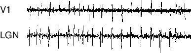
Fig. 2.8. Spontaneous activity in the cat PGO pathway. Each
line shows a 65second electrode recording from a cell in the indicated
area during REM sleep in the cat. Spontaneous REM sleep activation in
the pons of the brainstem is relayed to the LGN of the thalamus
(bottom), to the primary visual cortex (top), and to many other
regions in the cortex. It is not yet known what spatial patterns of
visual cortex activation are associated with this temporal activity,
or with other types of internally generated activity during
sleep. However, such activity is largely genetically determined and
could affect how the visual system develops. Reprinted with permission
from Marks et al. (1995), copyright 1995 by Elsevier.
|

