Click on the image to see a PDF version (for zooming in)
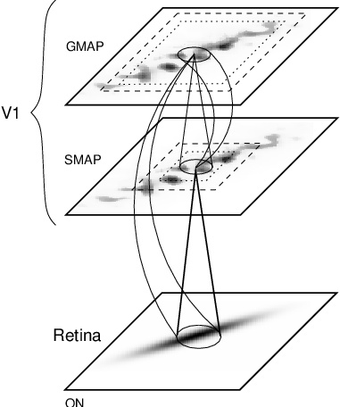
Fig. 11.1. Architecture of the PGLISSOM model. The cortical
network consists of two layers (or maps). The lower map (SMAP) has
short-range lateral excitation (dotted square) and long-range lateral
inhibition (dashed square), and drives the self-organization of the
model. In the upper map (GMAP), both excitation and inhibition have
very long range, establishing perceptual grouping and
segmentation. The two maps both receive afferent input directly from a
model retina, representing the ON channel like the reduced LISSOM
model (Figure 6.3). The neurons in the vertically corresponding
locations on the two maps are connected via excitatory intracolumnar
connections in both directions, tying such neurons together into a
functional unit (i.e. a cortical column). All neurons are spiking
neurons (Figure 11.2); their firing rate is visualized in gray-scale
coding from white to black (low to high).
|

