


Research
Quick links:: [Publications] [KESM specifications] [Gallery]
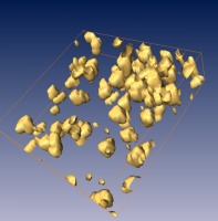
3D reconstruction of the hippocampus: A small portion of Nissl-stained hippocampus is visualized in 3D. [More images] [More nuggets] |
Our research team has expertise ranging from neuroanatomy, instrumentation, data acquisition, image processing, 3D graphics and visualization, cluster computing, to theoretical and computational neuroscience. The knife-edge scanning microscope (KESM) project involves aspects touched upon by all of the above areas.
Currently, our major focus is on the following tasks:
- Tissue staining: En bloc staining methods
Development of en bloc staining methods for whole mouse brains. Golgi, Nissl, and other stains. (Abbott).
[Gallery] - KESM instrumentation: KESM instrumentation and data acquisition
Sectioning and imaging of whole mouse brains under 100 hours. Knife chatter abatement. Tissue ribbon extraction. (McCormick, Gutierrez-Osuna, and Wiercigroch).
[Gallery]
[KESM specifications] - 3D reconstruction: Neuron morphology
Reconstruction of neuron cell bodies and processes from sequence of 2D images from KESM (Keyser and McCormick).
[Gallery] - Connectivity maps: Mouse brain web (MBW) distributed storage and network connectivity analysis
XML-based storage of neuron morphology and connectivity data in a web environment for scalable and efficient storage and retrieval. Development of network connectivity analysis algorithms for the study of natural computations in the mouse brain (Choe, Kim, and McCormick).
[Gallery] - Multi-scale integration: Reconstruction of mouse brain tissue bridging multi-scale imaging
From gross neuroanatomy (macro-scale brain atlas) through cellular structure (micro-scale light microscopy) to subcellular structure (nano-scale electron microscopy). Lab-wide effort, in collaboration with Stephen J. Smith at Stanford. (Mouse brain atlas data provided by Arthur Toga at UCLA Laboratory of Neuroimaging.)
Contact us | Home
© Copyright 2004 Brain Networks Laboratory | Department of Computer Science
Dwight Look College of Engineering | Texas A&M University | Texas Engineering Experiment Station

